:
JEOL JXA-8530F Field Emission Electron Probe Micro Analyzer

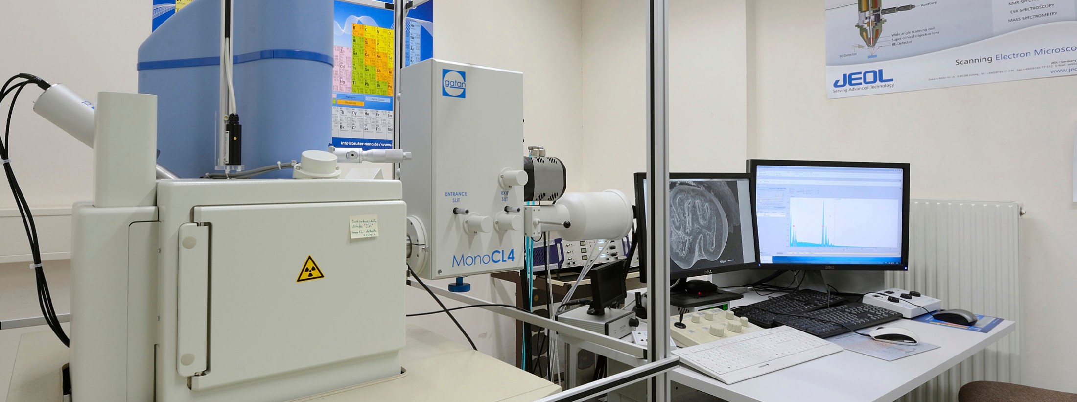
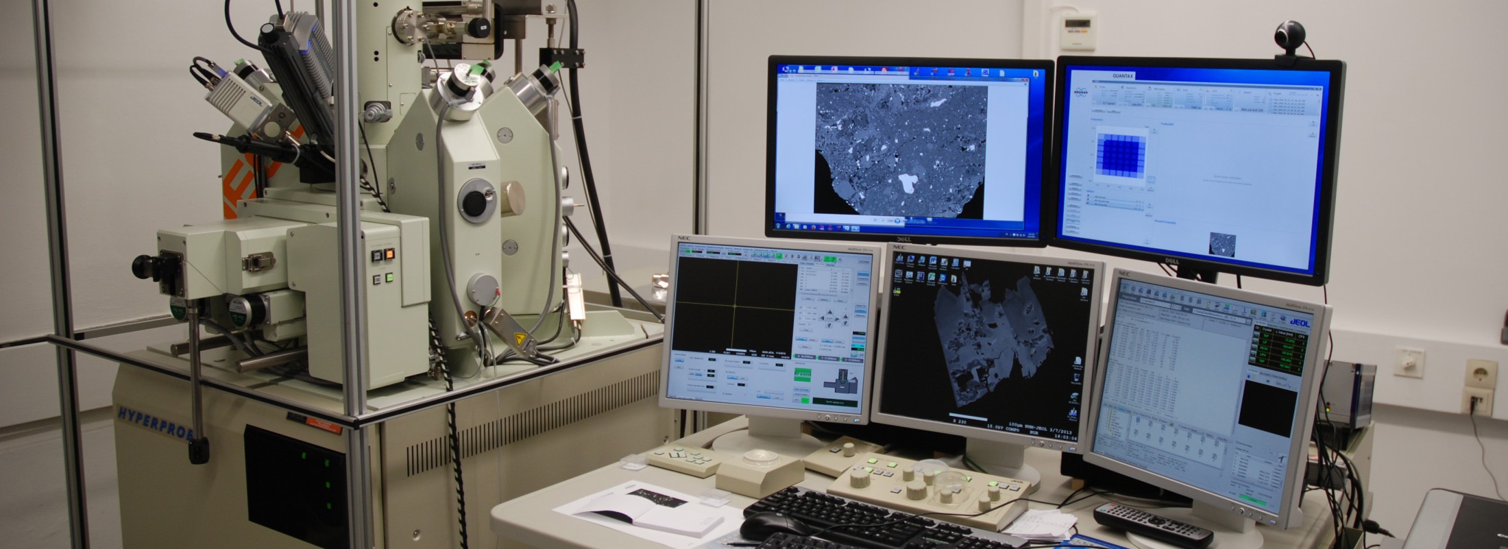
The JEOL JXA-8530F Hyperprobe is an electron probe micro analyzer with field emission cathode. The instrument is equipped with five WDS spectrometers and two EDS detectors. Elements from Be to U can routinely be analyzed in 0.01 wt% concentrations (in some cases even below 100 ppm) at a lateral resolution reaching into the sub-micrometer range. The integration of special image and analysis software packages combined with the fully automated state-of-the-art instrument offer the user unique analysis possibilities.

Vacuum system: oil rotary pumps, turbo-molecular pump, sputter ion pumps, differential pumping
Ultimate pressure: FE gun chamber ~ 10-8 Pa, sample chamber ~ 10-4 Pa
Electron gun: Schottky field emission electron gun
Accelerating voltage: 1 – 30 kV
Magnifications: ×40 – ×300,000 (WD 11 mm)
Probe current: 10 pA – 0.5 µA at 25 kV
Resolution: 3.0 nm at Acc. V. 30 kV, 10 pA, WD 8mm, SEI
Image output: 8-bit, up to 5120 × 3840 pixels
Specimen stage movement:  X 90 mm, Y 100mm, Z 7.5 mm
Specimen stage drive: minimum driving step size: X, Y: 0.02 µm/step, Z: 0.5 µm/step
Working distance: 11 mm
Integrated visible-light microscope: ½ inch CCD, reflected light, × 300, field of view 0.3×0.2 mm
Secondary electron (SE) detector: collector electrode, scintillator and photomultiplier tube
Backscattered electron (BSE) detector: Si P-N junction
Wavelength Dispersive X-ray Spectrometer (WDS) system:
  • five linear focusing type spectrometers with automatic analyzing crystal exchange at any point
  • X-ray take-off angle: 40°
  • Xe-filled and gas-flow proportional counters
  • seven analyzing crystals types: LDE1, LDE2, TAP, PETJ, PETH, LIF, LIFH
  • detectable wavelength: 0.087 – 9.3 nm
  • detectable/quantifiable element range: B – U
JEOL Energy Dispersive X-ray Spectrometer (EDS) system:
  • type: 10 mm2 Si(Li) semiconductor, Peltier cooling
  • detectable element range: B – U
  • quantifiable element range: Na – U
  • energy resolution: 138 eV or better (Fe, 5.9 KeV)
Computer automation: complete JEOL software package
  • WDS/EDS qualitative, quantitative analysis software
  • WDS/EDS line and mapping analysis software
Additional detector for EPMA:
  • Bruker XFlash 6/10 energy dispersive detector
Additional software for EPMA:
  • Bruker Esprit 2.1
  • Bruker Esprit 2.1 software
  • Probe for EPMA software
-  Multi-element area maps (beam scan and X-Y stage scan modes)
-  Matrix corrections include ZAF, CITZAF, PRZ, PROZA. Heinrich, Henke and/or
empirical mass absorption coefficients, calibration curve, thin film methods
-  Advanced background models for trace element analysis (Automated iterated   polynomial fit MAN (corrected for absorption of the continuum) or off-peak background corrections (linear interpolation, average or single side and exponential fits) or any combination of background corrections within a single sample)
- Quantitative spectral interference correction for both major and trace element analysis
- Integrated Area-Peak-Factor (APF) correction for light element analysis
- Quantitative graphical volatile element correction for any or all elements using both calibration reference, and/or internally referenced "self" calibration
  • Probe IMAGE software
  • Monte Carlo simulation software (Casino, Penempa, CalcZAF):
 to explore, for natural and theoretical matrixes and practical analytical conditions, the dimensions of excitation and escape volumes for different X-rays
  • Modal image analysis software
:
JEOL JSM-6610 LV Scanning Electron Microscope (SEM)

The JEOL JSM-6610 LV scanning electron microscope is a general-purpose instrument with a thermal tungsten cathode. Its low-vacuum capability allows high resolution imaging and chemical analysis of uncoated samples. A large sample chamber allows the investigation of up to dm-sized irregular shaped objects. Several attachments are currently in use: BRUKER energy dispersive system, BRUKER EBSD unit, GATAN cathodoluminescence system MonoCL4.
 
Vacuum system: oil rotary pump, oil diffusion pump, reservoir tank
Ultimate pressure: HV gun and sample chamber 0.1mPa: LV sample chamber 10-100Pa
Electron gun: pre-centered tungsten (W) hairpin filament
Accelerating voltage: 0.3 – 30 kV
Magnifications: ×5 – ×300,000
Probe current: 1 pA – 150 nA, at 20 kV
Resolution: 3.0 nm at Acc. V. 30kV, WD 8mm, SEI
Image output: 8-bit, up to 5120 × 3840 pixels
Specimen stage movement:  X 125mm, Y 100mm, Z 80 mm, Tilt -10 to 90º,
Rotation: endless
Attachments:
  • four-quadrant solid state backscattered (BSE) detector
  • variable pressure secondary electron (SE) detector
  • BRUKER energy dispersive X-ray detector (EDS)
  • BRUKER Electron Back-Scattered Diffraction (EBSD) detector
  • GATAN low magnification cathodoluminescence (Mini-CL) detector
  • GATAN cathodoluminescence system (MonoCL4)
Low-vacuum mode (variable pressure):
  • for imaging and qualitative X-ray analysis of uncoated material (type specimens)
  • best image resolution will be obtained at approximately 20-30 Pa
JEOL JCM 5000 Neoscope

The JEOL JCM 5000 Neoscope scanning electron microscope is a tabletop instrument with a thermal tungsten cathode. Its low-vacuum capability allows high resolution and fast imaging of uncoated samples. The scanning electron microscope is equipped with an SE detector for a magnification up to 40000x. The device is primarily intended for general presentation for museum visitors, however it is also used by scientific staff upon consultation.
:
Vacuum system: oil-rotary- and turbo-molecular pump
Electron gun: Small cartridge electron source
Accelerating voltage: 5, 10, 15 kV
Magnifications: ×10 – ×40,000
Image output: 1280 × 1024 pixels, bmp, tif, jpg
Specimen stage movement:  35 mm travel in X and Y
Sample size: Max.70 mm diameter and 50 mm thickness
Dimensions: 492x 458 x 434mm (W x D x H)
Features:
  • Automatic and manual control with pre-stored recipes
  • High and low vacuum modes
  • No special sample preparation, such as coating and drying, for conductive and non-conductive samples
  • Secondary electron and backscattered electron imaging
  • Image in three minutes after loading specimen
 
For questions, appointments and conditions, please contact: 
•Dr. Wencke Wegner ; P: 0043-1-52177-630
  
Online-Tickets