Analytics


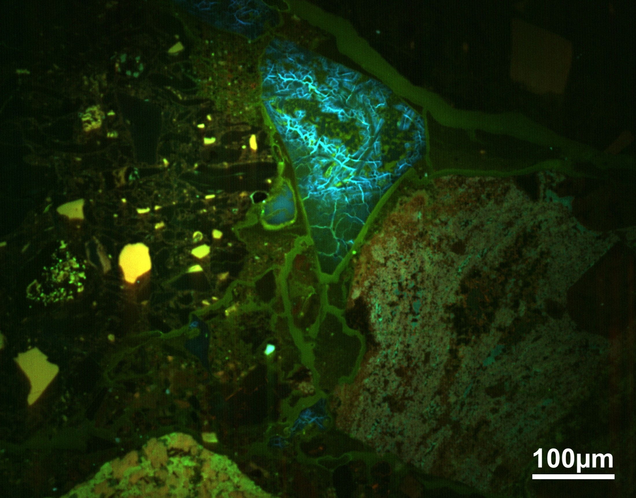
Backscattered electron (BSE), characteristic X-ray, and cathodoluminescence (CL) signals are used to get different information about the sample. X-rays are used for chemical characterization of the sample. CL and BSE are used to obtain structural and chemical information from the material analyzed.
 
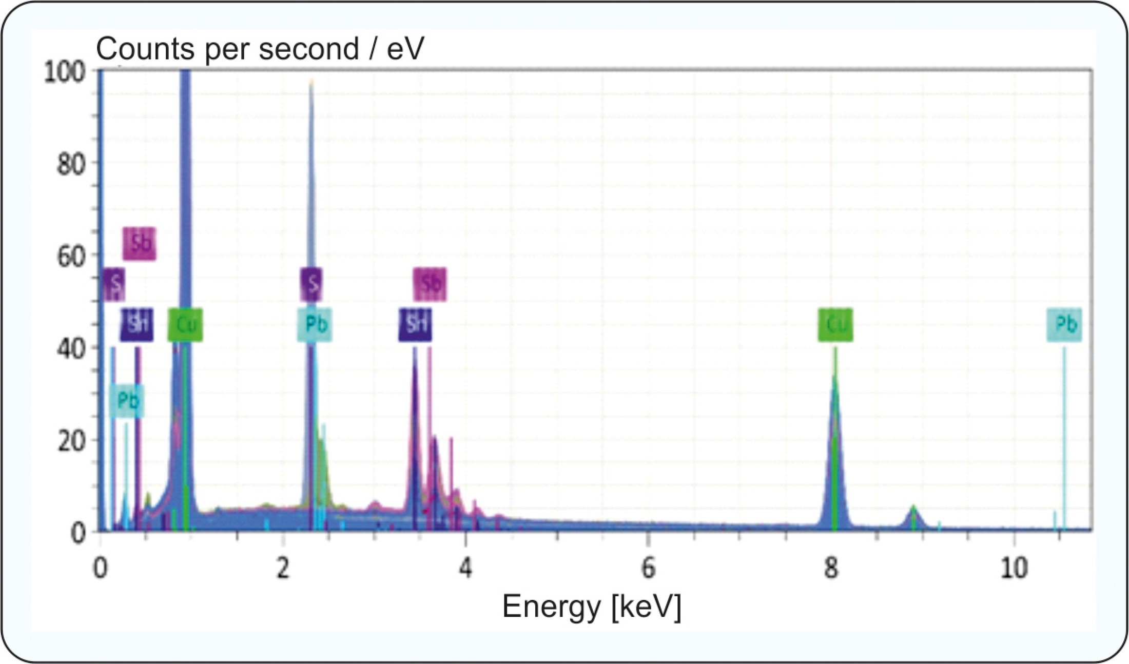
Energy-Dispersive Spectrometry (EDS)
The energy-dispersive spectrometry (EDS) is a method used for rapid identification of elements (qualitative analysis) and elemental abundances (quantitative analysis) in a point or an area of a solid sample, using a solid-state (energy-dispersive) detector. The detector collects and counts all of the emitted X-rays at once, sending the energy and number of a specific photon type into a proper energy channel. All X-ray energies from 0 to 25 keV are collected and displayed simultaneously as an energy spectrum in which the characteristic X-ray lines of the existing elements are displayed (section from an energy spectrum see figure).

:

The method can be used for phase identification, rapid particle analysis etc. For some combinations of elements, however, the method is affected by limited sensitivity and low energy resolution that leads to peak overlaps.
In this case, the higher-resolution WDS method must be used for reliable element identification.
Wavelength-Dispersive Spectrometry (WDS)
The wavelength-dispersive spectrometry (WDS) is a method used for accurate identification and measuring of the characteristic X-ray of interest. The higher resolution, compared to the EDS, is achieved by diffraction of X-rays on a crystal that is positioned between the sample and the detector. The so called analyzer crystal works as monochromator and only X-rays of a specific wavelength will be detected. Different analyzer crystals are used to cover the entire X-ray spectrum:
lithium fluoride (LIF), pentaerythritol (PET), thallium acid phthalate (TAP), and artificial layered dispersive element (LDE) crystals. Our EPMA has the complete selection of analyzer crystals, which are distributed over 5 spectrometers. This enables the simultaneous measurement of up to 5 elements.
WDS analysis results in a spectral resolution and sensitivity an order of magnitude better than obtainable with EDS analysis. The detection limits of WDS normally varies between 300 and 30 ppm. Compared to EDS, the WDS offers higher precision quantitative analyses, particularly for light elements, and better resolution of overlapping X-rays peaks to improve respectively enable element identification and quantification.
:
:
However, the user has to have an idea what kind of elements are in the sample as only elements are measured which are preselected by their wavelength. Therefore, a quick pre-check by EDS is recommended.
 
Cathodoluminescence
Cathodoluminescence (CL) is light or electromagnetic radiation (emitted from a sample), ranging from the ultraviolet to the near-infrared regime of the electromagnetic spectrum, which is generated by a high-energy electron beam. Electrons from the lower-energy ground state are promoted to the higher-energy excited state.

Composite RGB CL (Cathodoluminescence) image of some clasts in an impact brecia from the El’gygytgyn impact structure in Russia. The intensity of the luminescence is inversely correlated with the shock degree experienced by the investigated clasts.: Composite RGB CL (Cathodoluminescence) image of some clasts in an impact brecia from the El’gygytgyn impact structure in Russia. The intensity of the luminescence is inversely correlated with the shock degree experienced by the investigated clasts.
Composite RGB CL (Cathodoluminescence) image of some clasts in an impact brecia from the El’gygytgyn impact structure in Russia. The intensity of the luminescence is inversely correlated with the shock degree experienced by the investigated clasts.
When the energetic electrons attempt to return to the ground state, they may be temporarily trapped by intrinsic (structural defects) and/or extrinsic (impurities) factors in intermediate energy levels.
Energy is released by emitting a photon when high-energy electrons gradually decline to the ground state. The intensity of the CL is generally a function of the density of the intermediate energy levels. CL emissions can provide general information on the trace elements contained in substances or the production of mechanically induced defects in a material. The distribution of the CL in a material gives fundamental insights into such processes as crystal growth, element replacement, deformation, provenance and quality.

Electron Backscatter Diffraction (EBSD)
Electron backscatter diffraction (EBSD) is a method for the microstructural-crystallographic characterization of a crystalline or polycrystalline material. The incident primary electrons are scattered inelastically at the atoms of the sample, creating a divergent source in the sample. If some electrons hit lattice planes in such a way that the Bragg condition is fulfilled, constructive interference occurs. This reinforcement occurs for all lattice surfaces in the crystal, so that the resulting diffraction pattern, also called the Kikuchi band, shows all the angular relationships in the crystal and thus also the crystal symmetry. The method is used to structurally identify a phase, to find grain orientations and to study microstructures and deformations.


EBSD image of Cr3Si showing Kikuchi pattern (example image from Bruker), allowing the identification of crystal structure of the selected mineral phase.: EBSD image of Cr3Si showing Kikuchi pattern (example image from Bruker), allowing the identification of crystal structure of the selected mineral phase.
EBSD image of Cr3Si showing Kikuchi pattern (example image from Bruker), allowing the identification of crystal structure of the selected mineral phase.
  
Online-Tickets